Location
Mount Vernon, WA 98274

Location
Mount Vernon, WA 98274
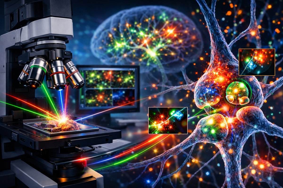
A breakthrough multicolor microscopy platform now captures live synaptic dynamics in mammalian brain tissue with unprecedented speed and resolution. This real-time imaging method promises to reshape our understanding of neural circuits, disease mechanisms, and the very essence of thought.
In a leap that brings the inner workings of the brain into vivid focus, researchers have unveiled a multicolor, volumetric imaging system capable of recording synaptic activity in real time across three dimensions. By combining advanced light-sheet illumination with multiphoton excitation and ultrafast detectors, this approach watches neurons speak to one another at the speed of thought. Early demonstrations in mouse hippocampal slices reveal wave-like calcium transients sweeping through dendritic spines and axon terminals, offering neuroscientists a direct window into learning and memory mechanisms that until now were largely inferred from static or slow recordings.
The heart of the new platform lies in a custom optical architecture that marries dual-color light-sheet modules with a branched two-photon pathway. Low-energy laser pulses excite genetically encoded calcium indicators deep within tissue, while a second wavelength simultaneously tracks voltage changes using faster fluorescent reporters. A series of microfabricated aspheric lenses and remote focusing mirrors sweep the light-sheet through a thick sample in fractions of a millisecond, all without mechanical stage movements. This combination reduces phototoxicity and avoids the motion artifacts that have long plagued live-tissue imaging.
Key to faithfully reporting rapid synaptic events are the genetically encoded indicators themselves. The team optimized a variant of GCaMP6f for brightness and response speed, pairing it with a red-shifted voltage sensor known as Voltron. When expressed in neurons, these probes glow brighter or dimmer in response to calcium influx and membrane depolarization, respectively. In head-to-tail tests, the dual sensors could resolve individual synaptic potentials and spontaneous action potentials even in congested circuits-information that was previously accessible only by invasive patch-clamp electrodes.
Handling the torrent of data generated by kilohertz-rate, three-dimensional scans required a matching leap in computation. Real-time processing pipelines harness graphics processing units to deconvolve raw images on the fly, reconstructing volumetric movies without delay. Machine-learning models trained on synthetic datasets correct for residual aberrations and assign each fluorescent flash to its precise spatial location. By the time a researcher glances at the monitor, entire networks of neurons are already rendered in color-coded activity maps, toggling on and off like constellations in a digital sky.
The implications for circuit neuroscience are profound. Investigators can now watch how groups of synapses co-activate during the formation of place fields in the hippocampus, or observe inhibitory and excitatory inputs interlocking to sculpt spike timing. In disease models, the platform illuminates how seizures originate from aberrant synchronization or how neurodegenerative processes erode communication at dendritic spines. Such direct visualization could accelerate the search for therapeutic targets by revealing pathological dynamics that were previously obscured.
Beyond fundamental research, this technology promises to transform translational studies and drug discovery. Pharmaceutical screening often relies on surrogate measures of neuronal health or indirect assays. By contrast, live-tissue imaging reveals the immediate effects of candidate compounds on synaptic physiology, allowing for rapid toxicology assessments and efficacy validation. Academic-industry collaborations are already underway to test thousands of small molecules in organotypic cultures, reducing the time from hit identification to lead optimization.
As with any powerful tool, ethical considerations surface alongside technical achievements. Animal welfare regulations guide the preparation of acute brain slices, and researchers adhere to the 3Rs-replacement, reduction, refinement-when designing experiments. Data privacy is emerging as a concern in large-scale brain initiatives, where imaging datasets could theoretically reveal individual-level characteristics if linked to behavioral records. Open-science advocates call for anonymized repositories and clear consent frameworks when human tissue or patient-derived organoids are imaged.
Long-term sustainability and resource use have also shaped the design. Engineers selected low-power laser diodes and recyclable optical components to minimize energy consumption and waste. Fluidic chambers that maintain tissue viability are machined from autoclavable plastics and glass, reducing the need for single-use microfluidic chips. Even the laboratory electronics are modular, so failed boards can be replaced without discarding entire systems. These choices align with growing calls for greener scientific infrastructure across research institutions.
Looking ahead, the team envisions integrating real-time optogenetic control with volumetric imaging, closing the loop between observation and manipulation. By stimulating targeted neurons based on observed activity patterns, researchers could probe causal relationships with millisecond precision. In parallel, efforts are underway to miniaturize the system into head-mounted form factors for small-animal studies, enabling behavioral experiments that map synaptic dynamics during navigation, social interaction, or decision-making.
Clinical translation remains a long-term horizon, but the path is already taking shape. Insights gleaned from tissue-slice experiments inform electrode design for deep-brain stimulation in epilepsy and movement disorders. Electrical engineers are adapting miniature imaging probes for intraoperative monitoring, allowing neurosurgeons to visualize synaptic integrity in real time. Meanwhile, computational frameworks developed for data deconvolution are being repurposed to analyze clinical electroencephalography and magnetoencephalography recordings, bridging scales from microscopic synapses to whole-brain rhythms.
“Seeing synapses light up in living tissue is more than a technical milestone,” says one of the lead investigators. “It changes how we ask questions about the brain. We can test hypotheses about network computations in ways that were purely theoretical until now.” The excitement reverberates through neuroscience departments around the world, as laboratories prepare to adopt this platform and push the boundaries of what’s observable.
In an era defined by big data and computational power, the ability to watch the fundamentals of neural communication in action represents both a scientific and philosophical advance. It invites us to reconsider the relationship between mind and matter, between thought and the biophysical events that underpin it. The hippocampal place cell that lights up when we enter a familiar room is no longer a black box; its flicker of calcium is now visible in living color. And with that visibility comes the potential to decode the language of consciousness itself, one synapse at a time.